SickKids researchers create a first-time, high-resolution image of key enzyme from the brain
Summary:
SickKids researchers have created a 3D model to allow scientists to better understand the V-ATPase enzyme's structure and how it functions.
This is a never-before-seen 3D video showing how a key piece of molecular machinery functions in order to keep many of the cells throughout your body working properly.
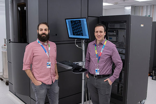
Researchers at The Hospital for Sick Children (SickKids) have created this detailed model to allow scientists to better understand the enzyme’s structure and how it functions. The study is published in the March 12 online edition of Science.
This enzyme, called V-ATPase, is found throughout the body and plays an important role in many diseases. V-ATPase in the brain is mutated in some forms of epilepsy and cancer. For the first time, we were able to see what the enzyme looks like in a mammal's brain.
John Rubinstein, Senior Scientist, Molecular Medicine
Previous research, also from the Rubinstein laboratory, looked at the enzyme in yeast cells but with the development of new methodology, the team was able to obtain and visualize the V-ATPase enzyme in mammals with an astonishing level of detail.

A number of steps had to take place in order to create the final model. First, Dr. Yazan Abbas, lead author of the study and Research Fellow in the Molecular Medicine program at SickKids, separates out the molecular structures the team wants to look at in detail.

These samples are prepared on tiny 3 mm grids, each one not much bigger than the head of a pin!


The grids are placed in a device that cleans them with plasma so the water-based solution that holds the samples can be applied evenly across the grid, ensuring the clearest picture possible.

The sample has to be flash-frozen to immobilize the V-ATPase for imaging in the microscope, but without allowing any ice crystals to grow, which could damage the sample.
With the sample spread on the grid, a robot blots the liquid into a thin film and plunges it into a mixture of liquid ethane and propane, which is kept at nearly -200 C with liquid nitrogen.

Now, the sample is ready for the microscope!

After extensive analysis of the images with state-of-the-art computers and software, the finished product is a blueprint of the V-ATPase. Researchers can use the blueprint to develop new drugs to effectively target the enzyme.

“Our lab will continue to develop new techniques in the hopes that we can understand how V-ATPase malfunction leads to disease and create precise interventions to treat the conditions that occur when it does go wrong,” says Rubinstein, who is also a Professor in the Departments of Biochemistry and Medical Biophysics at the University of Toronto.
This work was supported by the Canadian Institutes of Health Research, the European Research Council, Wellcome Trust, Restracomp and the Canadian Research Chairs Program.
CryoEM data was collected at the Toronto High-Resolution High-Throughput cryoEM facility, supported by the Canada Foundation for Innovation and Ontario Research Fund.

